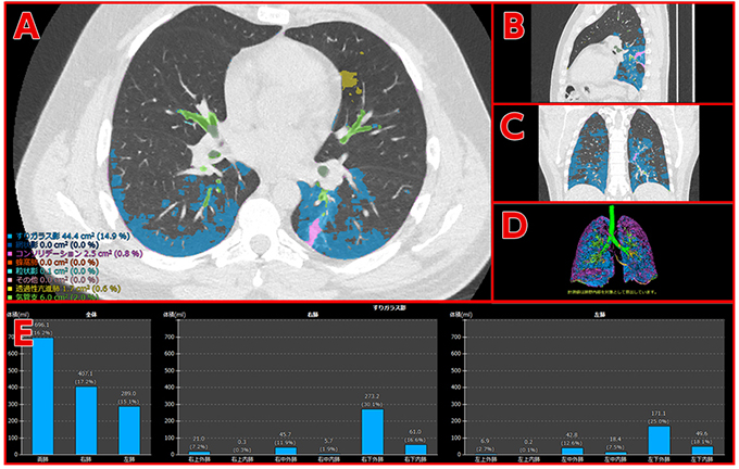
(Under development) CT images of a patient who has developed pneumonia as a complication of COVID-19 and analysis results A: CT image’s axial view (transverse plane), B: Sagittal view (longitudinal plane) C: Coronal view (vertical plane into ventral and dorsal, D: 3D image, each showing the results of lesion identification E: Graphs showing the presence of a specified type of lesions by zone and its volume
Fujifilm to Develop AI-based Technology to Aid COVID-19-Induced Pneumonia Diagnosis and Assess The Effectiveness of Treatments
Company commences joint research study with leading medical institutions in Japan, applying lesion quantification technology for pneumonia
TOKYO, May 19, 2020 — FUJIFILM Corporation (President: Kenji Sukeno) is commencing a research study to develop Artificial Intelligence (AI)-based technology to aid in the diagnosis and treatment assessment of patients with COVID-19-induced pneumonia. The technology for quantifying the lesions of interstitial pneumonia*, co-developed with Kyoto University (the Department of Respiratory Medicine, Graduate School of Medicine, Professor Toyohiro Hirai), will be applied to the project. The company will now embark on a joint research study with local medical institutions treating COVID-19 patients, starting with the Kanagawa Cardiovascular and Respiratory Center (Yokohama, Japan).
The spread of the novel coronavirus, which causes COVID-19, has emerged as a serious issue around the world. The world has yet to see clear judging criteria for determining the effectiveness of various treatment options, currently explored by doctors. In order to confirm the progression of pneumonia and the effectiveness of treatments, doctors need to examine hundreds of chest CT images for each patient to visually check the characteristics of ever-changing lesions and it puts a serious strain on specialists. There are expert opinions that COVID-19-induced pneumonia presents similarly to interstitial pneumonia in diagnostic images and has diverse variations in lesion patterns. Fujifilm’s CT quantification technology for interstitial pneumonia is powered by an AI-based software that examines CT images to identify bronchi, blood vessels and normal lungs in lung field** as well as seven types of lesions such as reticular opacities, ground-glass opacities and honeycomb lungs***, and automatically carries out categorization and measurement to quantify lesions of interstitial pneumonia. It also divides the lung field into 12 zones*4 and shows the volume and ratio of lesions for each of the zones so that clinicians can examine the distribution and progression of lesions within the lung field in details. Read More